
AI-Assisted Mammography for Breast Arterial Calcification and Cardiovascular Risk Prediction


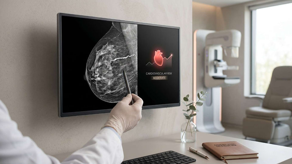
AI-assisted mammography is transforming how clinicians view the relationship between breast imaging and cardiovascular health. The presence of breast arterial calcification (BAC) on mammograms, once considered an incidental finding, is now emerging as a meaningful marker for cardiovascular risk prediction. Recent large-scale studies show that BAC provides a window into vascular aging and arterial stiffness—two critical factors linked to future heart disease and cardiovascular mortality. By leveraging artificial intelligence to quantify these calcifications, researchers are uncovering new ways to detect and prevent cardiovascular disease in women, especially those who may not yet show symptoms.
Understanding Breast Arterial Calcification and Its Clinical Significance
Breast arterial calcification refers to calcium deposits that build up within the arteries of the breast tissue. Unlike breast calcifications that might indicate early cancer or benign processes, BAC reflects calcium buildup in the arterial walls, similar to what occurs in coronary arteries. This process indicates vascular aging and may correspond to systemic arteriosclerosis, a condition that narrows and hardens the major arteries of the body. Identifying calcifications in the breast through routine mammography therefore provides more than information about breast cancer—it can also serve as a visual indicator of a woman’s cardiovascular health status.
How Common Is BAC in Mammography?
Breast arterial calcification is not rare. Studies suggest that mild BAC may be present in up to one in five women undergoing routine breast screening. The likelihood increases with age, diabetes, hypertension, and chronic kidney disease. However, many clinicians historically overlooked BAC because standard mammography interpretations focused primarily on cancer detection. The integration of AI-assisted mammography now helps quantify BAC more accurately and consistently, making it a valuable secondary metric in routine screening.
The Role of AI-Assisted Mammography in Quantifying BAC
AI-assisted mammography uses advanced machine learning algorithms to detect and measure the extent of breast arterial calcifications automatically. These algorithms identify calcium deposits and quantify them in square millimeters. By translating visual data into numerical values, AI creates standardized, reproducible results that radiologists and cardiologists can use for cardiovascular risk assessment.
How Does AI Quantify Breast Arterial Calcifications for Heart Risk Prediction?
In recent research from Emory University and the Mayo Clinic, AI quantified BAC and categorized findings as absent, mild (>0–10 mm²), moderate (>10–25 mm²), or severe (>25 mm²). This classification revealed a clear dose-response pattern: mild calcification increased the risk of cardiovascular events by about 30%, moderate by up to 70%, and severe BAC by as much as 200–300%. Over a seven-year observation period, women with visible calcifications were significantly more likely to experience heart attacks, strokes, or cardiovascular death than those without any signs of BAC.
AI-Based Screening for Cardiovascular Disease Prevention
Traditional cardiovascular screening often requires separate appointments, blood tests, or imaging studies such as coronary calcium scoring. However, AI-assisted mammography introduces the potential for dual screening. As millions of women undergo regular mammograms for breast cancer detection, AI could simultaneously evaluate cardiovascular risk without extra cost or radiation exposure. This dual-use approach could dramatically improve early identification and prevention of cardiovascular disease in women, a population often underdiagnosed or misclassified in conventional screening models.
Clinical Evidence Supporting BAC as a Predictor of Cardiovascular Mortality
The link between BAC and cardiovascular mortality is supported by large multi-site population studies. One such study analyzed more than 120,000 mammograms from women with no prior cardiovascular disease and found that BAC independently predicted future cardiovascular events even in women under 50. An editorial by Dr. Lori B. Daniels from the University of California, San Diego emphasized that breast arterial calcification is not just a benign imaging feature but a sign of vascular aging and arterial stiffness. She noted that including BAC quantification in mammogram reports could enhance awareness of cardiovascular risk in women who might otherwise be considered low-risk based on traditional factors.

Correlation Between BAC Severity and Cardiovascular Outcomes
Researchers identified strong correlations between the degree of BAC and cardiovascular outcomes. Women with severe BAC had the highest rates of major heart events, while those with mild or moderate levels also displayed elevated risk compared to women without any visible calcification. The data reinforces that BAC severity acts as a graded marker for cardiovascular health. Detecting and documenting even mild cases can therefore help prompt timely interventions, including lifestyle modifications and preventive treatment.
Integrating AI-Assisted Mammography Into Clinical Workflows
Bringing AI-based BAC quantification into clinical use requires thoughtful implementation. The first step is establishing standardized reporting methods that clearly differentiate cancer-related findings from vascular calcification. Automated systems can generate structured results that include BAC scoring and corresponding risk levels. Radiologists can then flag these findings for clinicians, prompting cardiovascular evaluation when warranted.
Policy and Practice Implications for Preventive Care
Integrating automated cardiovascular assessments into mammography workflows could shift the paradigm of preventive medicine. Policymakers and healthcare systems can consider incorporating BAC quantification into national mammography protocols, ensuring that women receive comprehensive risk assessments during routine screenings. Beyond clinical benefits, such integration promotes cost efficiency by using existing infrastructure for expanded health insights.
Addressing Communication and Ethical Considerations
While AI improves detection accuracy, clear communication of results remains essential. Patients should be informed about what BAC means, how it relates to their heart health, and whether further evaluation is necessary. Balanced communication avoids unnecessary anxiety yet empowers women to discuss prevention strategies with their healthcare providers. Experts also emphasize that BAC should be viewed as a marker of systemic health rather than a direct measure of coronary blockage.
Limitations and Future Research Directions
Despite the promise of AI-assisted mammography for BAC analysis, limitations exist. Not all types of calcifications correlate directly with coronary artery disease, and differences in imaging equipment or demographic factors may influence detection rates. Future research aims to refine algorithms for better accuracy across diverse populations and to conduct prospective clinical trials assessing how BAC-informed interventions impact long-term outcomes like cardiovascular mortality and heart disease prevention.
Technical Innovations Driving Progress
Machine learning and deep neural networks continue to advance the precision of image interpretation in mammography. As training datasets expand, AI systems will increasingly recognize subtle forms of vascular calcification and differentiate them from benign breast calcifications. Furthermore, integration with electronic health records can enable individualized cardiovascular risk stratification combining mammographic data with blood pressure, cholesterol, and lifestyle metrics.
Practical Applications for Cardiovascular Risk Stratification
Automated BAC scoring represents a significant opportunity for risk stratification in preventive cardiology. For example, women with moderate or severe BAC may benefit from more intensive monitoring, lipid management, and lifestyle interventions. Conversely, BAC absence could reassure patients with otherwise borderline risk factors. In this way, artificial intelligence in mammography enhances precision prevention by tailoring care based on objective imaging biomarkers.
Collaboration Between Radiology and Cardiology
AI-assisted mammography bridges the gap between two disciplines: radiology and cardiology. Radiologists can provide objective BAC data while cardiologists interpret its implications in the broader cardiovascular context. This collaboration fosters integrated care pathways where heart disease prediction is initiated from a simple mammogram rather than advanced cardiac testing.
Conclusion
AI-assisted mammography for detecting and quantifying breast arterial calcification has redefined how clinicians approach cardiovascular risk prediction. By transforming routine breast imaging into a dual-purpose diagnostic tool, artificial intelligence offers an accessible, cost-effective method to identify women at risk for heart disease and cardiovascular mortality. As research continues and healthcare systems adopt standardized BAC scoring, this innovation promises to enhance prevention strategies, improve early detection, and ultimately reduce the burden of cardiovascular disease in women worldwide. The future of screening is not just about detecting cancer—it is about protecting the heart as well.


Comments