
Breast Artery Calcification on Mammograms May Predict Heart Disease Risk in Women

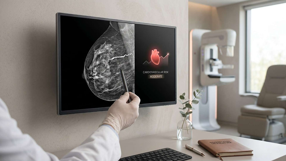
Routine mammograms have long been a cornerstone of women’s preventive health. Traditionally used to detect early signs of breast cancer, new research is revealing that mammograms may hold even more clinical value. The appearance of breast artery calcification (BAC) on these images could indicate future heart disease risk in women. Studies from institutions like Dartmouth College, Penn State, and others have shown a consistent link between breast arterial calcification and cardiovascular events such as heart attack and stroke. This discovery is changing how experts view mammogram imaging, suggesting that these common diagnostic tests could also become valuable tools for predicting cardiovascular risk.
What Is Breast Artery Calcification?
Breast artery calcification refers to calcium deposits within the arteries of the breast. Unlike cancer-related microcalcifications that appear in breast tissue, BAC occurs inside the blood vessels that supply the breast. These deposits are typically seen on mammograms as linear or track-like white lines. Researchers are now recognizing that this vascular calcification can reflect underlying systemic arterial stiffening, an early warning sign of cardiovascular disease. The findings suggest that BAC might not simply be a localized breast change but rather a marker of a broader vascular health issue, especially in aging and postmenopausal women.
How Mammograms Help Detect Cardiovascular Risk
When radiologists examine mammograms, they sometimes note the presence of calcium in breast arteries even though it is not currently part of standardized reporting. Recent evidence indicates that this simple observation could be clinically relevant. In the Dartmouth study involving over 1,200 women, BAC was detected in about 20 percent of participants. Those women were twice as likely to experience major cardiovascular events within 10 years compared to those without BAC. This correlation remained significant even when traditional risk factors were considered.
Can Routine Mammograms Predict Future Heart Disease in Women?
The evidence so far suggests they can. Women routinely undergo mammogram screening every one to two years after age 40. These existing images could offer insight into cardiovascular health without requiring additional testing or radiation exposure. If breast arterial calcification is noted, cardiologists could use this information to evaluate heart disease risk more accurately and take preventive action earlier.
Breast Artery Calcification vs. Traditional Cardiovascular Risk Models
The Pooled Cohort Equation, a widely used cardiovascular risk calculator, estimates a person’s 10-year risk of events like heart attacks and strokes. However, several studies indicate that it underestimates cardiovascular risk in women. In contrast, the presence of BAC appears to provide additional predictive power. For instance, among women who scored as low risk according to traditional equations, those with BAC still had a significantly higher chance of developing heart disease. Incorporating BAC data improved model accuracy and better identified which patients might benefit from lifestyle changes or preventive therapies.
Evidence from Clinical Research
A large Penn State analysis of more than 10,000 women confirmed these earlier findings. Participants with worsening BAC over four years had up to a 93 percent higher risk of heart attack, stroke, or death. This study also utilized artificial intelligence software, cmAngio AI, to automatically measure BAC severity levels—negative, mild, moderate, or severe. Such technology could make population-level cardiovascular screening more accessible using data already captured through mammography.
The Role of Breast Arterial Calcification in Postmenopausal Women
Another important study, focusing on women aged 60 to 79, found that BAC presence was independently associated with a 1.51-times higher risk of atherosclerotic cardiovascular disease. Even after adjusting for high blood pressure, diabetes, smoking, and other factors, BAC continued to predict elevated cardiovascular risk. The same research observed that only high levels of BAC strongly correlated with outcomes, suggesting a threshold effect. It emphasized that BAC should be formally recognized as a risk enhancer in preventive cardiology, especially among postmenopausal women.
Why BAC Matters After Menopause
After menopause, a decline in estrogen increases arterial stiffness and calcium buildup in blood vessels. These physiological changes make vascular health monitoring even more important for older women. Detecting BAC early could help clinicians identify postmenopausal women at high risk for cardiovascular events and allow targeted interventions aimed at reducing stroke and heart disease risk.
Should Radiologists Report Breast Calcification During Mammogram Screening?
Currently, radiologists may note breast calcification for breast cancer evaluation but are not required to report BAC findings. Dr. Thara Ali and other experts advocate for changing that approach. Reporting BAC, even informally, could provide valuable insights into a woman’s systemic vascular health. It may also encourage early discussion between patients and healthcare providers about addressing modifiable risk factors such as cholesterol, blood pressure, physical inactivity, smoking, and obesity. Including BAC in mammography reports could bridge a critical information gap between radiology and cardiology.
How Integrating BAC Reporting Could Enhance Preventive Care
If radiologists begin flagging significant calcium buildup in breast arteries, primary care physicians and cardiologists could include that data in cardiovascular risk calculators. This integrated view of breast and heart health might lead to earlier preventive measures, from prescribing statins to emphasizing diet and exercise strategies. Given the high mortality rate of heart disease in women, such proactive care could have a measurable public health impact.
Using Technology and AI to Advance Breast Calcification Detection
Emerging AI algorithms trained on digital mammography images can automatically detect and quantify calcification. This approach could make it possible to assess cardiovascular risk on a national scale without additional imaging costs. AI’s role in adjusting for variables such as age, breast density, and vascular patterns further enhances precision. Combining AI-based BAC detection with existing clinical data could create an efficient, low-cost cardiovascular screening method for women.
Potential Clinical Implementation Challenges
Despite promising data, researchers caution that incorporating BAC into clinical workflow still requires more study. One unresolved question involves standardizing how to quantify calcification and what thresholds should trigger warnings. There is also a need to understand whether BAC behaves similarly across ethnic groups and varying health backgrounds. Until larger, long-term studies confirm these patterns, medical societies are unlikely to include BAC reporting in official mammography guidelines.
Limitations and Research Gaps
While BAC detection shows potential as a noninvasive cardiovascular risk marker, several limitations remain. Most of the current studies include primarily non-Hispanic white participants, leaving uncertainty about generalizability. The timing and mechanism of calcification progression also remain unclear—some women developed worsening BAC within just one year. Moreover, not all calcification necessarily indicates disease progression; differentiating between stable and active vascular changes remains a research priority.
Next Steps in Research
Ongoing trials aim to determine whether reducing modifiable risk factors after BAC detection leads to fewer cardiovascular events. Researchers are also exploring whether calcium detection through mammograms might eventually complement or replace higher-radiation tests like coronary CT scans. Based on the available data, incorporating BAC into screening programs could provide a cost-effective approach for large-scale cardiovascular prevention.
Practical Implications for Women’s Heart Health
For women, especially those over 40, understanding the connection between breast calcification and cardiovascular disease is vital. Even though the presence of BAC does not mean active heart disease, it signals a need for detailed cardiovascular evaluation. Physicians might recommend using a cardiovascular risk calculator, blood tests, and lifestyle assessments to create a prevention plan. Early recognition of this link empowers women to take control of heart health long before symptoms appear.
Steps Women Can Take After a BAC Finding
1. Discuss BAC results with both a primary care doctor and cardiologist.
2. Undergo a comprehensive cardiovascular risk assessment.
3. Focus on controlling cholesterol, blood pressure, and blood sugar levels.
4. Maintain consistent exercise routines and heart-healthy nutrition.
5. Avoid smoking and manage stress through lifestyle or behavioral therapy.
These steps align with preventive cardiology goals and can significantly lower the risk of future heart attacks and strokes.
Conclusion
Breast artery calcification on mammograms offers a promising, accessible pathway for improving women’s heart health screening. The growing body of evidence supports BAC as an independent predictor of cardiovascular events, especially for postmenopausal women. While more research is needed to determine standardized methods for reporting and intervention, the potential is clear: mammograms could become powerful dual-purpose tools for detecting both breast cancer and cardiovascular risk. By recognizing calcification in breast arteries as a cardiovascular warning sign, clinicians and patients can work together to prevent life-threatening outcomes and improve long-term heart health in women.



Comments